


Radiographic diagnosis plays a crucial role in modern dentistry, giving dentists essential information that visual exams alone cannot provide. Dental radiographs, commonly known as X-rays, allow practitioners to visualize internal structures of teeth, bones, and surrounding tissues. This enables the detection of various oral health issues such as cavities, periodontal disease, impacted teeth, and bone loss.
There are several types of dental radiographs, including bitewing, periapical, panoramic, and cone-beam computed tomography (CBCT). Each of which serves specific diagnostic purposes.

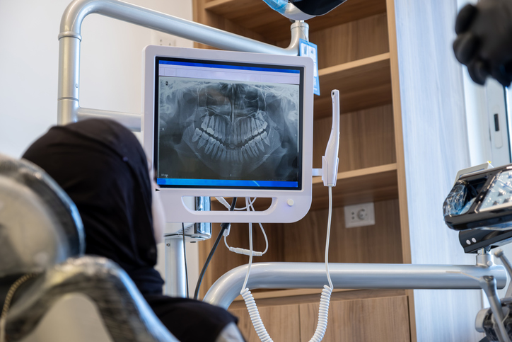
Radiographic Diagnosis in Your Exam
You’ll see us using different methods to capture pictures of your mouth, whether it’s during a comprehensive exam or cancer screening. We do this to acquire the most accurate results as possible.
Let’s make sure we don’t miss anything during your next visit. Call us at (515) 989-3180 or message us online to set up an appointment today!